13 Next steps
We’ve done a fairly streamlined preprocessing of a single cell spatial dataset. At this point in the analysis it would be time to drill into the biology and start doing some focussed analyses. What might be possible?
13.1 What did the authors find from this dataset?
In their paper Macrophage and neutrophil heterogeneity at single-cell spatial resolution in human inflammatory bowel disease Garrido-Trigo et al performed and extensive analysis of their cosmx Data alongside single cell, bulk and microscopy data. To summarise the kinds of things that are possible with single-cell spatial data - here is a list of some of the analyses from that paper that specifically use this spatial dataset;
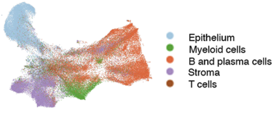
- Identified their detailed celltypes in their single cell data (over the whole transcriptome), which they used to classify celltypes in their spatial data. They classified their data at broad and specific levels (page 2 - Integration of single-cell RNA sequencing and spatial molecular imaging analysis provides a map of healthy and inflamed colon, Figs 1b and part of Supp Fig 1b for epithelia)
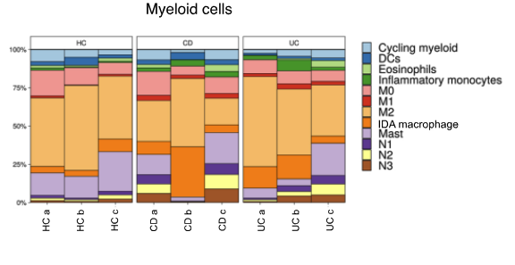
- Looked at differential abundance of celltypes across samples (page 2 - Transcriptional analysis at single-cell and spatial resolution reveals different populations of resident and inflammatory macrophages in the colonic mucosa )
- Identified cell niches with different neighbourhoods, e.g lymphoid structures, lamina propria and lower crypt. They quantified an increase in inflammatory celltypes in some of those neighbourhoods, and characterised another neighbourhood seen only in the IBD samples. (page 2 - Integration of single-cell RNA sequencing and spatial molecular imaging analysis provides a map of healthy and inflamed colon)
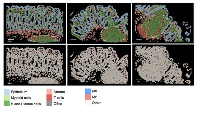
- They observed granuloma features in one patient. They examined the macrophage subtypes within them and the surrounding immune cell microenvironment. (Figure 4b, page 6 CosMx Spatial Molecular Imaging analysis confirms the expansion of Inflammation-Dependent Alternative macrophages and reveals their tissue distribution in inflammatory bowel disease colon)
- Identified a change in myeloid cell localisation by quantifying the distance of macrophage, MAST cells and Dendritic cells to the mucosal surface (page 2 - Transcriptional analysis at single-cell and spatial resolution reveals different populations of resident and inflammatory macrophages in the colonic mucosa )
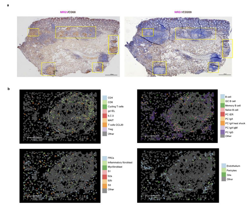
- Observed spatially localised expression of a gene (NRG1) within a subset of apically-located fibroblasts (page 4 - Inflammation-dependent alternative macrophages express neuregulin 1)
13.2 Other techniques
13.2.2 Downtream annotation and analyses
Niche analysis
Cell ‘niche’ analysis or ‘spatial context’ is a major avenue of in situ spatial analyses. E.g. tumour microenvrionments, tissue regions. The actual definition of what a ‘niche’ is could be considered flexible - it depends on what you are trying to achieve - and different tools take differnt approches.
- Seurat findNiches()
- OSCA neighbourhood analyses: https://lmweber.org/OSTA/pages/img-neighbourhood-analysis.html
- hoodscanR : https://www.bioconductor.org/packages/release/bioc/vignettes/hoodscanR/inst/doc/Quick_start.html
- GraphST : https://www.nature.com/articles/s41467-023-36796-3
- Many more …
‘Spatial’ pattern tests and cell free analyses
Huge area of research, here are some starting points;
- Co-localisation and differential co-localisation: https://lmweber.org/OSTA/pages/mult-diff-spatial-patterns.html
- Statial toolkit (Kontextual and Spatiomark) for quantifying spatial relationhips and expression with spatial contet: Statial package
- Spatial tests, adapted form geospatial methods: https://pachterlab.github.io/voyager/
- Molecule level data object (not a SingleCellExperiment): https://www.bioconductor.org/packages/release/bioc/html/MoleculeExperiment.html
- Many more …
Differential expression
Though current gen single-cell spatial techonlogies have lower counts and fewer genes in a panel than single cell (and bulk) studies, being able to test for differences in gene expression is valuable; potentially within a celltype, niche or spatial region that couldn’t be isolated with other technologies. With this in mind, single cell methods can be applied.
- Differential expression in scRNAseq (bioconductor): https://bioconductor.org/books/3.21/OSCA.multisample/multi-sample-comparisons.html
- Applied example (in this same dataset!) running differential expresison at the celltype level using Seurat or bioconductor ecosystem
- Spatially-aware differential expression methods; These are emerging quickly, consider looking into Differential gene expression analysis of spatial transcriptomic experiments using spatial mixed models and spacexr
- Many more …
Differential abundance
Differences in the numbers or types of cell in a particular location or neighbour niche between groups can indicate biological differences; from cell migration, expansion or development.
- Propeller for testing celltype proportion changes: https://github.com/phipsonlab/speckle
- Many more …
Multiomics
Another huge area of research, here are some starting points. It very much depends on which multiomics you have.
- Seurat multimodal vignette (single cell): https://satijalab.org/seurat/articles/multimodal_vignette (single cell)
- Image registration, for aligning images from different sources. E.g. H&E, immunofluorescence, proteomics e.t.c : https://lmweber.org/OSTA/pages/crs-spatial-registration.html
- Many more …
13.2.3 Visualisation
There are many tools out there, each with strengths and limitations.
- VRomics: https://ramialison-lab.github.io/pages/vromics.html
- Xenium explorer (Xenium only): https://www.10xgenomics.com/support/software/xenium-explorer/latest
- AtoMx (cosmx only) : https://nanostring.com/products/atomx-spatial-informatics-platform/atomx-sip-overview/
- Napari cosmx guide
- CellXGene (single cell) : https://cellxgene.cziscience.com/
- iSEE (single cell): https://github.com/iSEE/iSEE
- Many more …