Module 1.1: The omics landscape
Learning objectives
- Describe the molecular information captured by each of the five omics layers
- Explain how the molecular layers relate through the central dogma and its regulatory steps
- Select the most appropriate molecular layer for a given biological question
- Evaluate the limitations of each layer and what a chosen layer cannot tell you
Modern biology has undergone a fundamental shift from measuring one biological molecule at a time to profiling entire classes of biological molecules simultaneously. This lets us ask questions not just about individual genes, proteins, or metabolites, but about the state of a whole biological system, a tumour, a leaf under drought, a gut microbial community, at the molecular level.
Every living system, whether a bacterium, a migratory bird, or a human, can be interrogated across multiple molecular layers, and each layer reveals a different dimension of how the system works. The choice of which layer to investigate, and often which combination of layers, is one of the most consequential decisions a researcher makes before an experiment begins.
From DNA to metabolite
Most biological questions can be investigated at multiple molecular layers. Each molecular layer captures a different aspect of cellular biology. A biological question may be addressed at one layer or several, depending on what is actually driving the phenotype of interest.
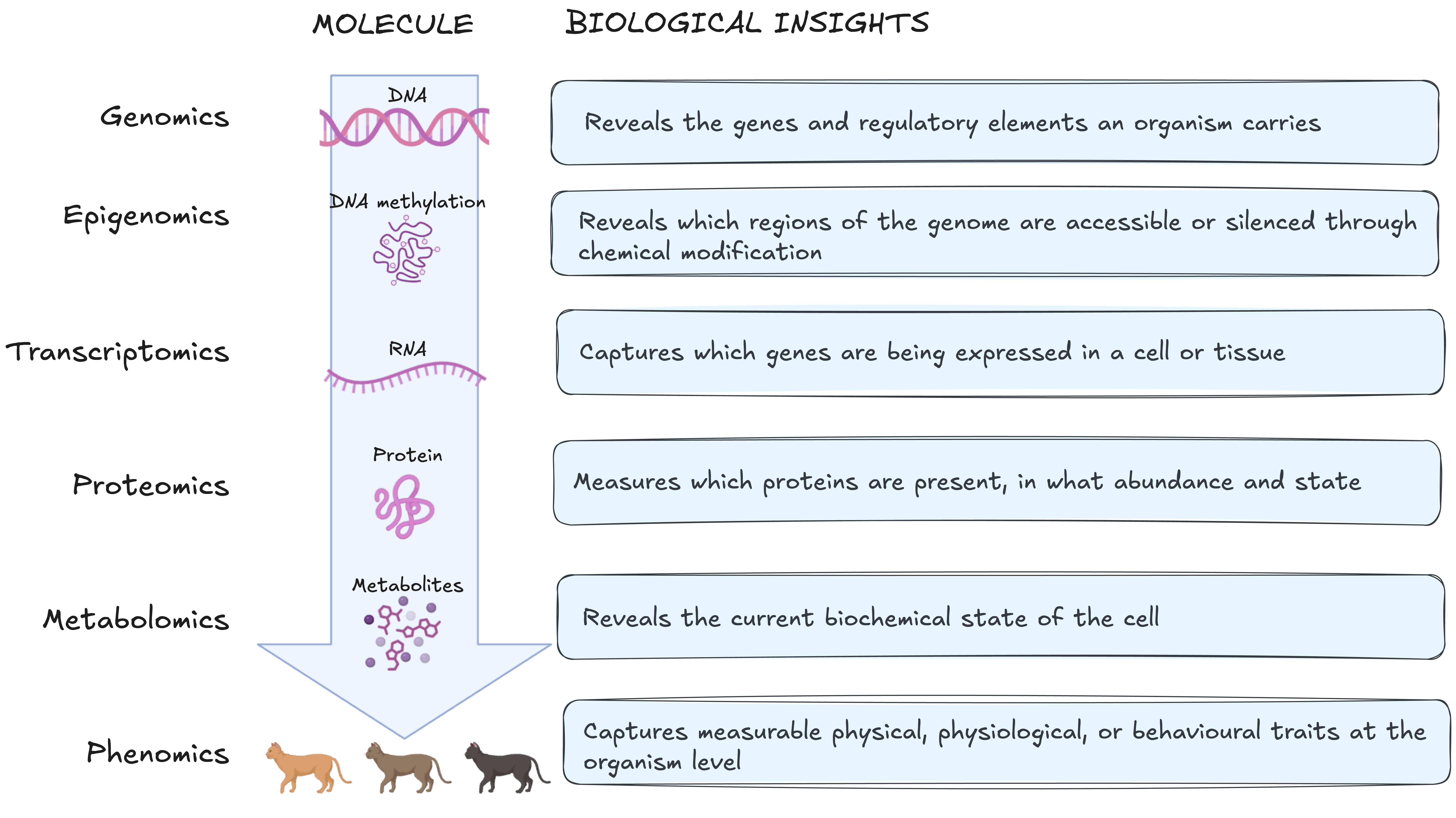
The molecular layers can be organised around the central dogma and its downstream and regulatory connections. The central dogma describes information flow from DNA to RNA to protein. Proteins then perform many cellular functions, including catalysing metabolic reactions. Metabolites and other cellular signals can also feed back to regulate these upstream layers. Exceptions and regulatory mechanisms complicate this picture but the framework remains a useful starting point for understanding how information flows between layers and where omics technology intervenes.
The figure below connects these five molecular layers through the central dogma and its regulation in the flow of biological information and what each layer captures.
Research study scenario: unexplained heart failure
A population of patients present to hospital with heart failure and the underlying cause is unclear. We will ask the same question at each molecular layer: what is driving these patients' heart failure?
These examples illustrate what each molecular layer could contribute. They do not imply that every sample type would be feasible or collected in a single study.
Layers overlap: the choice is fit, not exclusivity
Most questions can be approached from more than one layer. The lists below show what each layer is best suited to answer, not what it alone can answer. For example, if we want to know how closely two species are related, we can compare their DNA, RNA, or protein sequences. DNA is usually the best starting point because the same genome can be studied from any tissue, and it contains both coding and non-coding regions.
With RNA, we only capture genes that are expressed in the tissue we sampled. The general rule: pick the layer where your signal is the dominant source of variation, not one where it competes with variation you are not studying.
The order of layers traces information from genomic variation through to its molecular consequences. It is a conceptual model, not a one way pathway, the layers feed back on each other throughout.
Layer 1: DNA (the genome)
What is it?
DNA contains the instructions required to build and maintain cells. The genome is the complete set of DNA in an organism.
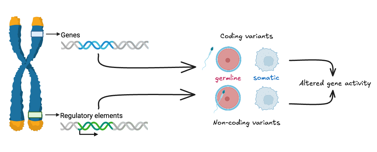
Most of the genome does not encode proteins. Non-coding regions include regulatory elements like promoters, enhancers, and silencers, that control when, where, and how a gene is transcribed. Many genes contain exons, which are retained in mature RNA, and introns, which are removed from pre-mRNA during RNA processing. Exons may contain protein-coding sequence or untranslated regions. The structure of a gene determines which RNA or protein isoforms can be produced from it and therefore which downstream layers are affected.
Understanding the genome is a prerequisite for interpreting the epigenome (which regions are regulated) and the transcriptome (which isoforms are expressed).
Role in biology
The genome is a stable repository of hereditary information. It determines which genes an organism possesses, how those genes are structured, and contains the regulatory elements that control gene activity across all downstream molecular layers. Unlike the layers beyond it, the inherited genome is relatively stable across a lifetime, although somatic mutations and other genomic changes can arise in particular cells or tissues.
Insights this layer can provide
Genomics can reveal which genes an organism carries, how they are structured, and what variants are present. This includes single nucleotide variants or polymorphisms (SNV/SNP), small insertions and deletions (indels), structural variants, and copy number changes. Variants may be germline, inherited and present in every cell, or somatic, acquired in specific tissues as in cancer. The non-coding genome is equally informative: variants in promoters, enhancers, and silencers can alter when and where genes are active without changing the protein sequence itself, and are increasingly recognised as important drivers of phenotypic variation.
Beyond individual biology, the genome supports comparative questions. Genomic variation is the basis for reconstructing evolutionary relationships, characterising genetic diversity within and between populations, and identifying signatures of selection. These applications span species identification, population genetics, conservation biology, and the study of how genetic diversity shapes disease susceptibility across groups.
What it can't tell us
The genome describes what an organism could do, not what it is doing.
A gene's presence tells us nothing about whether it is transcribed, how much protein it produces, whether that protein is active, or what metabolic consequences follow. Two individuals can carry the same variant and present with completely different phenotypes, because gene expression, epigenetic regulation, environment, and chance all mediate the path from genotype to phenotype.
The genome in our research question
Blood derived DNA from our heart failure patients can be screened for inherited variants in genes with established roles in cardiomyopathy. Whole genome or whole exome sequencing can identify rare pathogenic variants. For example, a truncating mutation in TTN (which encodes titin, the largest protein in the sarcomere and a major structural component of cardiac muscle) or a missense variant in MYH7 (beta myosin heavy chain, the primary motor protein driving cardiac contraction). Variants in these genes are among the most common monogenic causes of dilated cardiomyopathy.
- What we learned: a subset of patients carry pathogenic variants in sarcomere genes, providing a potential molecular explanation for their cardiac dysfunction.
- What we still don't know: whether those variants are penetrant in these individuals not all carriers develop disease and whether patients without identified variants have a different underlying cause that genomics alone cannot reveal.
Layer 2: DNA modification (the epigenome)
What is it?
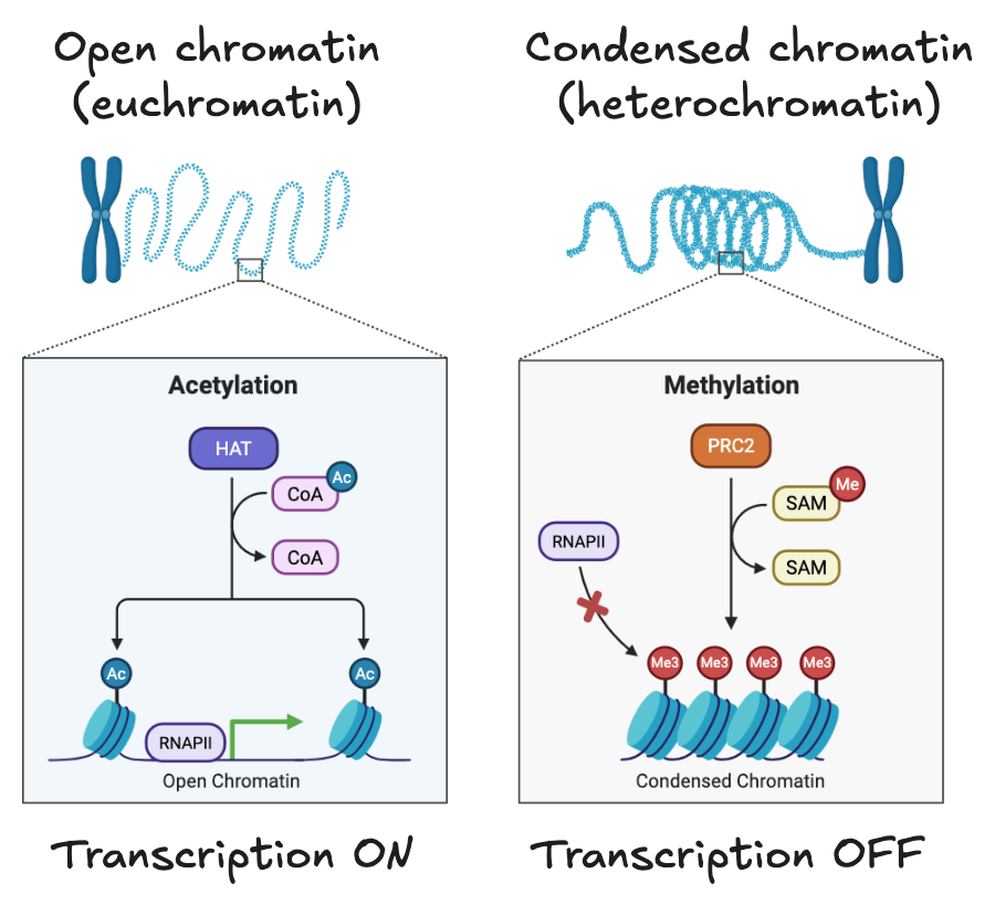
The epigenome includes chemical modifications to DNA and histone proteins, together with changes in how accessible the DNA is. These features influence how genes are regulated without changing the DNA sequence. Two major mechanisms contribute to this:
- DNA methylation: at gene promoters, it is associated with reduced transcription.
- Histone modifications: different modifications are associated with more open or compact chromatin and can influence transcription.
These modifications regulate gene activity without altering the underlying DNA sequence. The epigenome explains a fundamental puzzle in cell biology: how can a skin cell and a neuron contain identical DNA yet perform completely different functions? The answer lies in systematic epigenetic differences between cell types, which influence which genes are accessible and which are less accessible for transcription.
Role in biology
The epigenome acts as the regulatory interface between an organism's fixed genetic sequence and its dynamic environment. Developmental cues, ageing, and environmental exposures, including diet, stress, and toxins, can alter epigenetic marks, changing which genes are available for transcription without changing what those genes encode. The epigenome is therefore the layer at which genetic potential meets environmental context.
Insights this layer can provide
Epigenomics reveals the regulatory state of the genome in a given cell type at a given time. By measuring DNA methylation, histone modifications and chromatin accessibility, we can identify regulatory regions associated with greater or lower potential for gene expression information that DNA sequence alone cannot provide.
This is particularly valuable for understanding how the same genome produces different cell types during development, how environmental exposures alter gene regulation over time, and how disease states involve changes in chromatin accessibility rather than changes in sequence. Epigenomic data also helps interpret non-coding variants identified by genomics:
Epigenomic data can show whether a regulatory region containing a SNP is accessible or carries regulatory marks in the tissue of interest. This helps assess whether the SNP could affect gene regulation, although it does not prove that the SNP is functional.
What it can't tell us
Epigenetic changes indicate regulatory potential, not gene expression. An accessible chromatin region means a gene is available for transcription, not that it is being transcribed. Measuring DNA methylation or histone marks tells us nothing about whether accessible genes are actively producing RNA, how much, or in which isoforms. The epigenome also does not directly reveal whether altered regulation changes RNA abundance or downstream cellular function; these questions require evidence from the transcriptome and, potentially, downstream layers.
The epigenome in our research question
The epigenome can tell us which genes containing variants of interest are accessible for transcription in the failing heart. Profiling chromatin accessibility (ATAC-seq) or DNA methylation in cardiac tissue can reveal whether stress response gene regions have opened up. This is a pattern associated with cardiac remodelling under sustained pressure or volume overload. Epigenetic changes can reflect both pre-existing regulatory states and responses to disease or environmental conditions.
- What we learned: stress response and remodelling gene regions have become accessible in the patients' failing myocardium, consistent with active transcriptional reprogramming under sustained cardiac stress.
- What we still don't know: whether those accessible regions are actually being transcribed, and whether the epigenetic changes are driving disease progression, compensating for it, or both.
Layer 3: RNA (the transcriptome)
What is it?
The transcriptome is the set of RNA molecules present in a cell or tissue at a given time. Their abundance reflects both RNA production and RNA stability. Where the genome tells us which genes exist and the epigenome provides information about their regulatory state, the transcriptome tells us which RNA molecules are present and their abundance.
It provides one of the first molecular readouts of the cell's current state rather than its genetic potential.
The transcriptome captures more than which transcripts are present and at what abundance. Alternative splicing, which is the process by which different combinations of exons are joined during RNA processing, means a single gene can produce multiple distinct transcripts, called isoforms. Each isoform potentially encodes a protein with a different structure or function. Two samples with identical gene level expression can therefore differ substantially at the isoform level, with functional consequences that gene level analysis would miss.
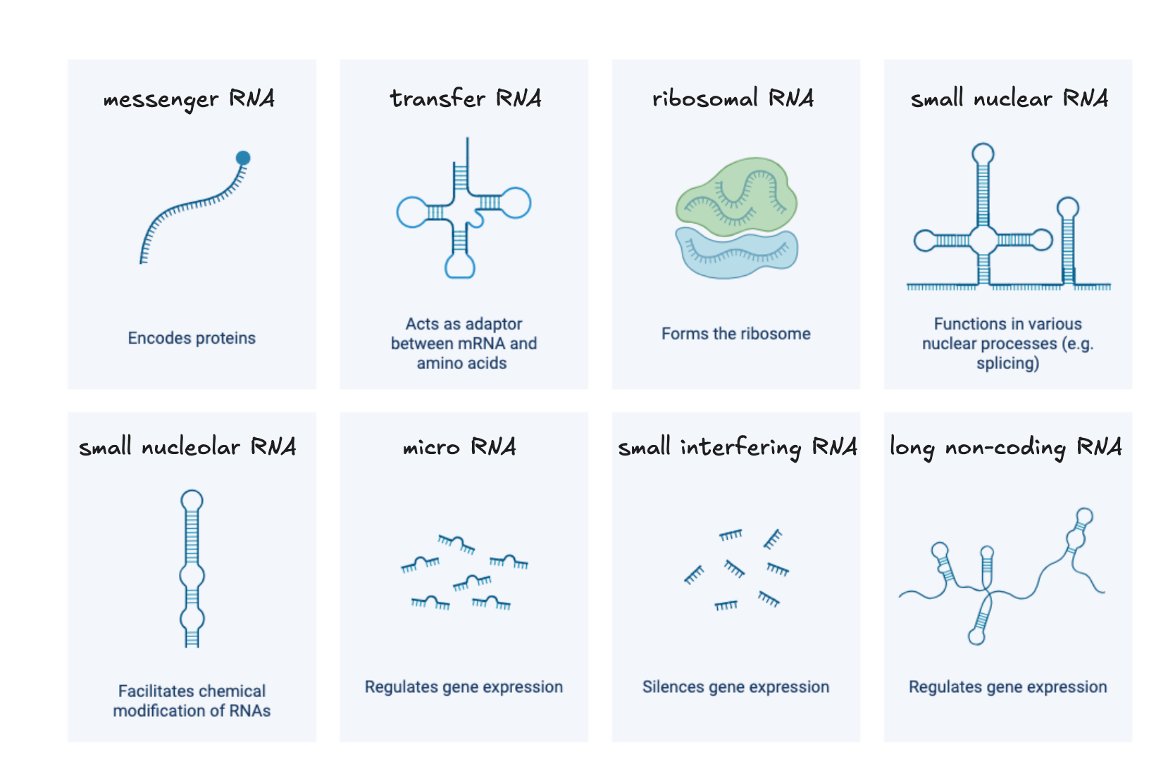
Beyond messenger RNA (mRNA), the transcriptome includes non-coding RNAs like microRNAs and long non-coding RNAs, that do not encode proteins but regulate gene expression, chromatin state, and RNA stability. Structural RNAs such as ribosomal and transfer RNAs are also transcribed constituents of the transcriptome, though they are typically removed in standard transcriptome studies. The regulatory non-coding RNA fraction is large, incompletely characterised, and increasingly recognised as central to the control of gene expression.
Role in biology
The transcriptome is the highly dynamic of the molecular layers. Gene expression changes rapidly in response to developmental signals, environmental conditions, disease, and treatment. This responsiveness makes it a sensitive readout of cellular state. But it also means results depend heavily on when and from which tissue the sample was collected. A transcriptomic snapshot captures one moment in a continuous, context dependent process.
Insights this layer can provide
Transcriptomics identifies which genes are active in a given cell or tissue, at what level, and in which isoforms. These are questions the genome and epigenome cannot answer directly. Differential expression (DE) analysis between conditions, e.g. disease versus healthy tissue or treated versus untreated cells, can reveal which pathways are involved and how the cell has reorganised its transcriptional programme in response. Because expression changes rapidly, transcriptomics is also well suited to capturing dynamic processes like responses to acute stress, progression through a developmental stage, or the early effects of a drug.
At the isoform level, transcriptomics can detect alternative splicing events that produce functionally distinct protein variants from the same gene. This is relevant in conditions where splicing is disrupted.
Transcriptomic profiling at single cell resolution adds a further dimension, revealing how gene expression varies between individual cells within the same tissue and enabling the identification of rare cell populations or transitional states that bulk measurements would obscure.
What it can't tell us
RNA abundance does not reliably predict protein abundance. Post-transcriptional regulation, including RNA stability, translational efficiency, and protein degradation rates, means that transcript and protein levels can diverge substantially. A highly expressed gene is not necessarily producing abundant or active protein, and a gene with low transcript levels may still maintain significant protein levels due to slow protein turnover.
The transcriptome in our research question
Moving from the epigenome to the transcriptome takes us from accessibility to observed activity. Measuring the transcriptome of failing cardiac tissue tells us which genes are up or downregulated relative to healthy myocardium, which signalling pathways, fibrosis, inflammation, hypertrophy, are engaged, and which isoforms are being produced from genes such as TTN, where isoform switching between the compliant fetal N2BA isoform and the adult N2B isoform has direct mechanical consequences for cardiac function.
- What we learned: stress-response and remodelling genes are actively transcribed; fibrosis and hypertrophy pathways are upregulated; isoform shifts in structural genes are detectable and functionally relevant.
- What we still don't know: whether those transcripts are being translated into functional protein at the expected levels, and whether the resulting proteins are correctly localised and active within the sarcomere.
Layer 4: Proteins (the proteome)
What is it?
Proteins are the major functional molecules of the cell. They catalyse the biochemical reactions that sustain life, form the structural scaffolds of cells and tissues, transmit signals, transport molecules, and regulate gene expression. The proteome is the complete set of proteins present in a cell, tissue, or organism at a given time.
Proteins rarely act in isolation. Many assemble into multi-protein complexes that are molecular machines whose activity depends on which subunits are present and in what relative proportions. The composition of these complexes can determine substrate specificity, regulatory sensitivity, and subcellular localisation in ways that measuring individual protein abundance cannot capture. A protein can be present at normal levels while its binding partners are absent, leaving the complex non-functional.
The relationship between a protein's amino acid sequence and its three-dimensional structure, and therefore its function, is not always predictable from sequence alone. Small sequence differences can produce large structural and functional changes, and post-translational modifications further alter how a protein folds, where it localises, and what it binds.
Role in biology
Proteins execute virtually every cellular function. Unlike RNA, which reflects transcriptional activity, the proteome provides a closer view of the molecules involved in cellular functions: which enzymes are present, which signalling proteins are altered, and which structural components are affected. The proteome integrates the effects of post-translational modification by phosphorylation, ubiquitination, acetylation, and others, that rapidly alter protein activity, localisation, and stability in response to cellular signals without any change in transcript levels. This regulatory layer is invisible to transcriptomics and only partially visible to genomics, making the proteome essential for understanding how cells respond dynamically to their environment.
Insights this layer can provide
Proteomics can identify which proteins are present and estimate their abundance. Specialised approaches can also measure post-translational modifications, protein interactions and subcellular localisation. In clinical contexts, proteins measurable in accessible biofluids such as plasma or urine serve as biomarkers of tissue-level pathology, reflecting changes in distant tissues that cannot be directly sampled.
Proteomics also reveals discordance with the transcriptome. A transcript can be upregulated while its protein product is rapidly degraded, or a protein can accumulate without a corresponding increase in its mRNA due to changes in translation efficiency or protein stability. These mismatches are biologically meaningful and would be missed by transcriptomics alone. For questions about what the cell is actually doing, the proteome provides evidence that no upstream layer can substitute for.
What it can't tell us
Protein abundance alone does not capture activity. A protein can be present in abundance while sequestered in the wrong compartment, held in an inactive conformation by an inhibitor, or absent from its functional complex. Post-translational modifications modulate activity in ways that standard abundance measurements may not detect without modification-specific enrichment strategies. The proteome also does not directly reveal the downstream metabolic consequences of protein activity — for that, the metabolome is needed.
The proteome in our research question
Measuring the proteome of cardiac tissue or plasma brings us close to the functional molecules involed in disease. Clinically informative biomarkers can be detected in plasma: e.g. elevated BNP and troponin in plasma can reflect cardiac stress and injury. Specialised phosphoproteomic or spatial approaches could investigate abnormal phosphorylation or localisation of contractile proteins. Proteomics can also expose mismatches with the transcriptomic picture, a transcript upregulated in the failing heart whose protein product is simultaneously being degraded, or a structural protein present at normal abundance but carrying modifications that impair its function.
- What we learned: BNP and troponin are elevated in plasma, and the abundance of several cardiac and inflammatory proteins differs from the comparison group. Specialised analyses also identify altered phosphorylation of contractile proteins.
- What we still don't know: what those protein level changes mean for the heart's real-time metabolic and energetic state, the question the next layer is positioned to answer.
Layer 5: Metabolites (the metabolome)
What is it?
Metabolites are small molecules produced, consumed, or modified during cellular metabolism.
They include sugars, amino acids, lipids, nucleotides, and organic acids - the substrates and products of the enzymatic reactions that sustain cellular life. The metabolome is the complete set of these molecules present in a cell, tissue, or organism at a given time.
Metabolites occupy a distinctive position in the molecular hierarchy. Where upstream layers describe genetic potential, regulatory state, RNA abundance and cellular machinery present, the metabolome provide a close readout of biochemical and physiological state at the moment of measurement. However, metabolite concentrations alone do not establish pathway activity or metabolic flux.
Beyond their role as metabolic intermediates, many metabolites function as signalling molecules, linking metabolic state back to gene regulation and completing a regulatory loop that runs in both directions through the molecular hierarchy.
Role in biology
The metabolome integrates information from all upstream molecular layers and from the external environment simultaneously. Nutritional state, oxygen availability, drug exposure, physical activity, microbial activity, and cellular stress all leave measurable signatures in the metabolome. This makes metabolomics a sensitive readout of whole-organism physiological state. It also means the metabolome reflects many influences at once, and attributing a metabolic change to a specific upstream cause requires supporting evidence from other layers.
Insights this layer can provide
Metabolomics directly measures the biochemical state of a cell or tissue at the time of sampling. It can reveal changes in metabolites and provide evidence of altered metabolic pathways and energetic state. Where these metabolites can be measured reliably, ratios such as ATP:ADP or NAD⁺:NADH can provide information about cellular energetic status. In disease contexts, characteristic metabolic signatures can serve as biomarkers of pathological state, and in pharmacology, metabolomics captures how a drug alters cellular biochemistry beyond its intended target.
Metabolomics is also the layer that closes the loop between molecular measurements and observable phenotype. The functional consequences of genetic variants, epigenetic changes, altered gene expression, and protein dysfunction ultimately manifest as changes in metabolic output. A metabolic shift observable in plasma or tissue is therefore often the most direct molecular correlate of a clinical phenotype — even when the upstream cause remains unclear.
What it can't tell us
The metabolome captures current state, not cause. A metabolic signature tells us what is happening now, not what initiated it. Establishing causality requires integrating evidence from upstream layers. Metabolites are also highly dynamic: concentrations can shift within minutes, and results are sensitive to pre-analytical variables including the time of sample collection, handling, freeze-thaw cycles, and the subject's nutritional state in the hours before sampling. Without rigorous standardisation of collection and processing, technical variation can obscure or mimic biological signal. Finally, the metabolome does not distinguish whether an observed metabolic change is a driver of pathology or a consequence of it.
The metabolome in our research question
Measuring the metabolome gives us the failing heart's real-time biochemical state. Heart failure is associated with metabolic remodelling, including changes in substrate utilisation, mitochondrial function, and energy production. Changes in the relative contribution of fatty-acid, glucose, and other substrates can occur during heart failure, but the direction and extent of these changes depend on disease stage, heart-failure phenotype, and metabolic context.
These metabolic changes can be reflected in tissue or plasma metabolite profiles, including changes in fatty-acid–related metabolites, TCA-cycle intermediates, lactate, ketone bodies, and other metabolites involved in energy metabolism. Together, these patterns can provide evidence of altered metabolic and energetic state, but metabolite concentrations alone do not establish pathway flux or whether a metabolic change is a cause or consequence of heart failure.
-
What we learned: the failing hearts show metabolic changes consistent with altered substrate utilisation and mitochondrial energy metabolism.
-
What we still don't know: whether these metabolic changes are driving disease progression or are a consequence of structural and functional changes in the failing heart and which upstream molecular changes contribute to them.
Summarising the layers
No single layer answered the question of what is driving these patients' heart failure. Each layer captured a different aspect of the underlying biology and left questions that could be addressed by integrating evidence from other layers.
| Layer | What it tells us | What it misses | Phenotypic question addressed |
|---|---|---|---|
| Genome | Genetic variation and potential | Whether variants affect gene function | Which genetic variants are associated with the phenotype? |
| Epigenome | Regulatory state | Whether genes are expressed | Which regulatory states are associated with the phenotype? |
| Transcriptome | Gene expression and isoform diversity | Whether proteins are produced and active | Which genes/isoforms are differentially expressed? |
| Proteome | Functional molecules and their state | Physiological consequences | Which proteins and protein states are altered? |
| Metabolome | Current physiological state | The underlying cause | Which metabolic processes and physiological states are altered? |
These layers are interconnected and feed back on each other. The table sets out where each layer is strongest, not a strict division of what each can address in isolation.
Activity
Each of the five molecular layers described above is studied by its own omics field: genomics, epigenomics, transcriptomics, proteomics and metabolomics. Omics approaches measure many features from a molecular layer simultaneously, providing a broad, system-level view. However, every platform captures only part of that layer, and omics studies may use either broad discovery-based or more focused targeted approaches.
Walk the layers
Pick the broad question closest to your field, or reinterpret an example using an organism or system you work with. These examples represent high-level biological questions rather than specific studies, and the same design principles apply across systems.
Walk through the molecular layers and decide how you would approach the question.
In your group, discuss:
- Which molecular layer would you start with, and why?
- What would this layer tell you, and what would it miss?
- What comparisons or timing would be needed to make the study meaningful?
Report back: Your chosen layer, what another layer might have revealed, and one assumption behind your choice.
Clinical / human disease
What drives progression from a treatable tumour to one that resists therapy?
Consider where you would sample, when in the disease course, and what each molecular layer would contribute to understanding progression.
Wildlife / infectious disease
Why do some populations tolerate an infectious disease while others experience severe disease from the same pathogen?
Consider whether this is a host question, such as immune response or genetic resistance; a pathogen question, such as strain or virulence factors; or both. You can use any host–pathogen system relevant to your field.
Aquaculture / production biology
Why do some farmed fish grow faster than others despite receiving the same diet?
Consider what might explain the variation if diet is held constant—for example, genetics, developmental history, physiology or gut microbes. Which layer would you measure first, and how would you sample to make the comparison meaningful?
Plant / environmental stress
How does a crop plant respond to acute environmental stress, and what makes some varieties more tolerant than others?
Consider whether you want to investigate the immediate stress response, the mechanisms associated with tolerance, or inherited differences between varieties. Which molecular layer and sampling times would best address your question?